The extracellular matrix (ECM) provides microenvironment for developing tissues and organs, by storing cytokines and growth factors and forming their gradient. Among molecules of ECM, proteoglycans play a major role in morphogenesis and organogenesis. Here, preparation of versican, a representative proteoglycan of ECM, and observations of their effects on oraganogenesis are described. |
| Category | Matrices & cellular trafficking |
| Protocol Name | Effects of the extracellular matrix on organogenesis |
Authors
 |
Watanabe, Hideto
Institute for Molecular Science of Medicine, Aichi Medical University
|
| KeyWords |
|
Reagents
 |
| ● |
4 M guanidine hydrochloride/ phosphate buffered saline (pH 7.2) |
| ● |
PMSF (100 mM in ethanol, working concentration = 1 mM) |
| ● |
N-ethylmaleimide (100 mM in water) |
| ● |
EDTA (0.5 M)
[Note] In place of proteinase inhibitors, proteinase inhibitor tablets (Roche Applied Science, Penzberg, Germany) can be used. Since versican is sensitive to proteinases, the solutions in each step should contain adequate levels of proteinase inhibitors. |
| ● |
|
| ● |
Chondroitinase ABC (Seikagaku Corp., Tokyo, Japan) |
| ● |
Bovine testicular hyaluronidase (Sigma-Aldrich, St. Louis, MO) |
| ● |
|
| ● |
Rabbit polyclonal anti-mouse versican CS-alfa and CS-beta (Merck Millipore, Billerica, MA) |
| ● |
Mouse monoclonal anti-CS antibody LY111 (Seikagaku Corp.) |
| ● |
Biotinylated hyaluronan (Seikagaku Corp.) |
|
Instruments
 |
| ● |
Ultracentrifuge (Beckman Coulter, Inc. Brea, CA) |
| ● |
Dot blot apparatus (ATTO Corporation, Tokyo, Japan) |
| ● |
Superose 6 column (GE Healthcare, Little Chalfont, UK) |
| ● |
DEAE-Sepharose (GE Healthcare) |
|
| Methods |
|
1. |
Preparation of versican from aorta*1 |
| 1) |
Mince bovine aorta* into small pieces. Wash the sample three times with PBS, and briefly centrifuge. |
Comment 1
|

|
| 2) |
Add ten volumes of 4 M GuHCl, and extract by rotating the sample overnight at 4°C. |
Comment 0
|
|
|
| 3) |
Centrifuge at 4,000 × g, 8°C for 30 min, and collect the supernatant as the 1st extract. |
Comment 0
|
|
|
| 4) |
Add five volumes of 4 M GuHCl to the precipitate, and extract by rotating the sample for 12 h at 4°C. |
Comment 0
|
|
|
| 5) |
Centrifuge at 4,000 × g, 8°C for 30 min, and collect the supernatant as the 2nd extract. |
Comment 0
|
|
|
| 6) |
Mix the 1st and 2nd extracts*, and dialyze against 50 mM Tris-HCl, pH 7.5, 7 M urea. |
Comment 1
|
|
|
| 7) |
Apply the sample to a DEAE Sepharose column (~5 mL) equilibrated in the same buffer, and wash with at least five column volumes of 50 mM Tris-HCl, 0.2 M NaCl, 7 M urea. Then, elute with 50 mM Tris-HCl, pH 7.5, 2 M NaCl, 7 M urea. |
Comment 0
|
|
|
| 8) |
Collect the eluate and dialyze against 50 mM Tris-HCl, pH 7.5. |
Comment 0
|
|
|
| 9) |
Add three volumes of 95% ethanol, 1.3% potassium acetate. Rest at −20°C overnight, and centrifuge at 10,000 × g for 30 min. |
Comment 0
|
|
|
| 10) |
Decant supernatant, and resuspend the precipitate in a small volume of 50 mM Tris-HCl, pH 7.5, 7 M urea. |
Comment 0
|
|
|
| 11) |
Apply the sample to a Superose 6 column. Monitor versican by dot blot analysis using anti-versican antibody. Collect the sample containing versican. |
Comment 0
|
|
|
| 12) |
Dialyze the sample against an appropriate buffer. |
Comment 0
|
|
|
|
2. |
Isolation of versican from aggrecan |
| 1) |
Extract proteoglycans from tissues using 4 M GuHCl as above. |
Comment 0
|
|
|
| 2) |
Add cesium chloride powder to give an initial density of 1.61 g/mL. |
Comment 0
|
|
|
| 3) |
Ultracentrifuge at 35,000 × g for 72 h. |
Comment 0
|
|
|
| 4) |
Fractionate the sample from the bottom. |
Comment 0
|
|
|
| 5) |
Perform dot blot analysis to detect versican and aggrecan. |
Comment 0
|
|
|
| 6) |
Collect versican rich fraction, and adjust the density to 1.6 g/mL, and ultracentrifuge at 35,000 × g for 72 h. |
Comment 0
|
|
|
| 7) |
Fractionate and perform dot blot analysis, collect versican rich fractions. |
Comment 0
|
|
|
| 8) |
Adjust the density to 1.51 g/mL, and ultracentrifuge at 35,000 × g for 72 h. |
Comment 0
|
|
|
| 9) |
Fractionate and perform dot blot analysis, collect versican rich fractions, and make sure that the sample no more contains aggrecan. |
Comment 0
|
|
|
|
3. |
[Cell culture experiments using versican, by treatment with chondroitinase ABC, and hyaluronidase*2] Treatment of cultured cells with chondroitinase ABC or hyaluronidase*3 |
| 1) |
Prepare the cells for analysis in as appropriate size. |
Comment 0
|
|
|
| 2) |
Replace the medium to a minimal volume of fresh one. |
Comment 0
|
|
|
| 3) |
Add chondroitinase ABC (5 milliunits/mL) or bovine testicular hyaluronidase (0.2–2 mg/mL), and culture for hours to days. |
Comment 0
|
|
|
| 4) |
Perform assay to investigate the role of CS. |
Comment 0
|
|
|
|
4. |
[Cell culture experiments using versican, by treatment with chondroitinase ABC, and hyaluronidase*2] Treatment cultured cells with versican |
| 1) |
Prepare the cells for analysis in an appropriate size. |
Comment 0
|
|
|
| 2) |
Replace the medium to a minimal volume of fresh one. |
Comment 0
|
|
|
| 3) |
Add versican isolated as above (final conc. 1–100 μg/mL), and culture (hours to days). |
Comment 0
|
|
|
|
5. |
[Cell culture experiments using versican, by treatment with chondroitinase ABC, and hyaluronidase*2] Immunostaining for versican, CS, and detection of hyaluronan*4 |
| 1) |
Plate cells onto LabTech chambers, and culture. |
Comment 0
|
|
|
| 2) |
Fix the cells with 4% paraformaldehyde for 30 min. |
Comment 0
|
|
|
| 3) |
Rinse the chamber slide with PBS. |
Comment 0
|
|
|
| 4) |
Treat with blocking solution for 30 min, and rinse the chamber slides briefly. |
Comment 0
|
|
|
| 5) |
Treat with (1) anti-versican CS-alfa and anti-CS-beta (1:1000) for 1 h, or (2) anti-CS LY111 (1:200) overnight at 4°C, or (3) biotinylated hyaluronan (2 μg/mL) for 15 min. |
Comment 0
|
|
|
|
|
| Notes | *1 GuHCl is used for extraction of proteoglycans. However, DEAE Sepharose column chromatography does not work in the presence of GuHCl. To obtain clear elution profiles, the buffers for column chromatography should contain 7 M urea. To precipitate proteoglycans using ethanol solution, urea should be removed by dialysis. Contaminant urea is precipitated by ethanol precipitation.
*2 In various assays using cell culture systems, we can investigate the regulatory role of the ECM on cell behavior, by manipulating the ECM molecules. Here, experiments using versican, chondroitinase ABC, and hyaluronidase, are described.
*3 Although bovine testicular hyaluronidase contains chondroitinase activity, we have observed that CS remains in cell culture. Thus treatment for a short period, its chondroitinase activity can be ignored.
*4 Methods to detect versican, CS and hyaluronan in mouse cell culture systems. |
| Figure & Legends |
Figure & Legends 
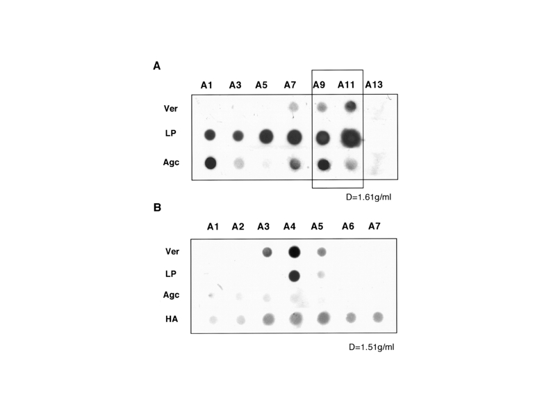
Fig. 1. Versican aggregate in normal articular cartilage
Dot blots of versican (Ver), link protein (LP), aggrecan, and hyaluronan (HA) are shown. A, patterns of initial density gradient ultracentrifugation (d, 1.61 g/mL) under associative conditions. B, patterns of density gradient ultracentrifugation (d, 1.51 g/mL) of fractions A9-A10 of the initial ultracentrifugation under associative conditions.
This figure was originally published in J Biol Chem. Matsumoto K, Watanabe H.et al. "Identification and characterization of versican/PG-M aggregates in cartilage" 2006, 281(26):18257–63. © the American Society for Biochemistry and Molecular Biology. |
| Copyrights |
 Attribution-Non-Commercial Share Alike Attribution-Non-Commercial Share Alike
This work is released underCreative Commons licenses
|
| Date of registration:2015-07-06 14:02:27 |
- Suwan, K., Choocheep, K, Hatano, S., Kongtawelert, P., Kimata, K., and Watanabe, H. (2009) Versican/PG-M assembles hyaluronan into extracellular matrix and inhibits CD44-mediated signaling toward premature senescence in embryonic fibroblasts. J Biol Chem. 284, 8596–8604 [PMID : 19164294]
- Matsumoto, K., Kamiya, N., Suwan, K., Atsumi, F., Shimizu, K., Shinomura, T., Yamada, Y., Kimata, K., and Watanabe, H. (2006) Identification and characterization of versican/PG-M aggregates in cartilage. J Biol Chem. 281, 18257–18263 [PMID : 16648631]
- Kamiya, N., Watanabe, H., Habuchi, H., Takagi, H., Shinomura, T., Shimizu, K., and Kimata, K. (2006) Versican/PG-M regulates chondrogenesis as an extracellular matrix molecule crucial for mesenchymal condensation. J Biol Chem. 281, 2390–2400 [PMID : 16257955]
|
This work is licensed under Creative Commons Attribution-Non-Commercial Share Alike. Please include the following citation
How to Cite this Work in an article:
Watanabe, Hideto,
(2015). GlycoPOD https://jcggdb.jp/GlycoPOD.
Web.25,4,2024 .
How to Cite this Work in Website:
Watanabe, Hideto,
(2015).
Effects of the extracellular matrix on organogenesis.
Retrieved 25,4,2024 ,
from https://jcggdb.jp/GlycoPOD/protocolShow.action?nodeId=t23.
html source
Watanabe, Hideto,
(2015).
<b>Effects of the extracellular matrix on organogenesis</b>.
Retrieved 4 25,2024 ,
from <a href="https://jcggdb.jp/GlycoPOD/protocolShow.action?nodeId=t23" target="_blank">https://jcggdb.jp/GlycoPOD/protocolShow.action?nodeId=t23</a>.
Including references that appeared in the References tab in your work is
much appreciated.
For those who wish to reuse the figures/tables, please contact JCGGDB
management office (jcggdb-ml@aist.go.jp).
|
|
