The concentration of heparan sulfate (HS) can be determined by enzyme-linked immunosorbent assay (ELISA). HS chains immobilized on the surface of Heparin/GAG Binding Plates are detected by an anti-HS antibody. Competitive ELISA is also a practical method for the quantitative estimation of HS concentrations. An anti-HS antibody is preincubated with increasing concentrations of standard HS, and the mixture is added to the biotinylated HS-immobilized plate. It should be noted that HS preparations derived from various sources differs in their reactivity to an anti-HS antibody depending on the epitope content along the HS chain. Only when the same HS preparation is used as a standard, can the concentration of HS be determined. To quantify uncharacterized HS preparations, ELISA methods might not be suitable, but HPLC after digestion with heparin lyases appears to be effective. |
| Category | Glycosaminoglycans |
| Protocol Name | Application of anti-GAG antibody and biotinylated hyaluronan binding protein(bHABP) [2]
~ Determination of the concentration of heparan sulfate using ELISA |
Authors
 |
Yamada, Shuhei
Department of Pathobiochemistry, Faculty of Pharmacy, Meijo University
|
| KeyWords |
|
Reagents
 |
| ● |
HS from bovine kidney (Seikagaku Corp., Tokyo, Japan) or porcine intestine (Sigma-Aldrich, St. Louis, MO)
Biotinylated HS |
| ● |
0.1 M Phosphate-buffered saline (PBS) |
| ● |
PBS containing 0.05% Tween-20 (PBST) |
| ● |
25 mM Tris buffered saline (TBS) |
| ● |
TBS containing 0.05% Tween-20 (TBST) |
| ● |
Bovine serum albumin (BSA) |
| ● |
Anti-HS antibody, F58-10E4 or HepSS-1 (Seikagaku Corp.) |
| ● |
The secondary antibody, anti-mouse IgG+IgM (H+L) conjugated with alkaline phosphatase (Kirkegaard & Perry Laboratories, Inc., Gaithersburg, MD) |
| ● |
2 mg/ml p-Nitrophenyl phosphate (pNPP) in 50 mM carbonate buffer, pH 9.8, containing 0.5 mM MgCl2 |
|
Instruments
 |
| ● |
Streptavidin Plate C8 Transparent (Nunc, Roskilde, Denmark) |
| ● |
Heparin/GAG Binding Plates (Iduron Ltd., Manchester, UK) |
| ● |
Microplate reader (Bio-Rad Laboratories, Hercules, CA) |
| ● |
|
|
| Methods |
|
1. |
ELISA using Heparin/GAG Binding Plates |
| 1) |
Wash the wells with 200 μL of PBS. |
Comment 0
|

|
| 2) |
Add HS/50 μL of PBS to the wells and incubate at room temperature overnight. |
Comment 0
|
|
|
| 3) |
Wash the wells with 200 μL of PBS. |
Comment 0
|
|
|
| 4) |
Add 200 μL of 1% BSA/PBS for blocking and incubate at 37˚C for 1.5 h. |
Comment 0
|
|
|
| 5) |
Wash the wells with 200 μL of PBST three times. |
Comment 0
|
|
|
| 6) |
Add 50 μL of the anti-HS antibody (diluted 1:150 in PBS) and incubate at 37˚C for 1 h. |
Comment 1
|
|
|
| 7) |
Wash the wells with 200 μL of TBST three times. |
Comment 0
|
|
|
| 8) |
Add 50 μL of the secondary antibody (diluted 1:2,000 in TBS) and incubate at 37˚C for 1 h. |
Comment 1
|
|
|
| 9) |
Wash the wells with 200 μL of TBST three times. |
Comment 0
|
|
|
| 10) |
Add 50 μL of the pNPP solution and incubate at room temperature. |
Comment 1
|
|
|
| 11) |
Measure the absorbance at 415 nm with a microplate reader (Fig. 1). |
Comment 0
|
|
|
|
2. |
Competition ELISA using streptavidin-coated plates |
| 1) |
Wash the wells with 200 μL of PBS. |
Comment 0
|
|
|
| 2) |
Add 2 μg each of biotinylated HS/50 μL of PBS to the wells and incubate at 4˚C overnight. |
Comment 1
|
|
|
| 3) |
Add 200 μL of 3% BSA/PBS for blocking and incubate at room temperature for 1 h. |
Comment 0
|
|
|
| 4) |
Incubate the anti-HS antibody (diluted 1:150 in PBS) and various concentration of HS in 50 μL of PBS in a test tube at room temperature for 30 min. |
Comment 0
|
|
|
| 5) |
Wash the wells with 200 μL of PBST three times. |
Comment 0
|
|
|
| 6) |
Add 50 μL of the anti-HS antibody or the mixture of the anti-HS antibody and HS to the wells. |
Comment 1
|
|
|
| 7) |
Incubate the plate at 37˚C for 1 h. |
Comment 0
|
|
|
| 8) |
Wash the wells with 200 μL of TBST three times. |
Comment 0
|
|
|
| 9) |
Add 50 μL of the secondary antibody (diluted 1:2,000 in TBS) and incubate at 37˚C for 1 h. |
Comment 1
|
|
|
| 10) |
Wash the wells with 200 μL of TBST three times. |
Comment 0
|
|
|
| 11) |
Add 50 μL of the pNPP solution and incubate at room temperature. |
Comment 1
|
|
|
| 12) |
Measure the absorbance at 415 nm with a microplate reader (Fig. 2). |
Comment 0
|
|
|
| Figure & Legends |
Figure & Legends

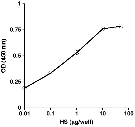
Fig. 1. The anti-HS antibody F58-10E4 interacts with HS in a dose-dependent manner.
Various concentrations of bovine kidney HS were immobilized on a Heparin/GAG Binding Plate from Iduron, and the binding was detected using F58-10E4, and then alkaline phosphatase-conjugated anti-mouse IgG/IgM secondary antibody. pNPP was incubated for 60 min, and the values represent the mean (n = 3).
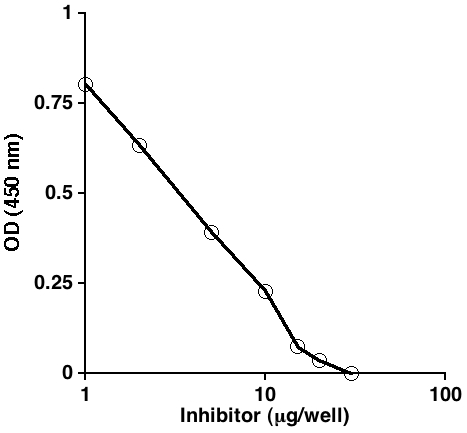
Fig. 2. The interaction of the anti-HS antibody F58-10E4 with biotinylated HS immobilized on a streptavidin-coated plate was inhibited by HS in a dose-dependent manner.
Biotinylated porcine intestinal HS was immobilized on the plate, and F58-10E4 preincubated with various concentrations of unlabeled porcine intestinal HS was added. The binding of the anti-HS antibody was detected with an alkaline phosphatase-conjugated anti-mouse IgG/IgM secondary antibody. pNPP was incubated for 60 min, and the values represent the mean (n = 2). |
| Copyrights |
 Attribution-Non-Commercial Share Alike Attribution-Non-Commercial Share Alike
This work is released underCreative Commons licenses
|
| Date of registration:2017-01-23 15:52:31 |
- Kure, S., and Yoshie, O. (1986) A syngeneic monoclonal antibody to murine Meth-A sarcoma (HepSS-1) recognizes heparan sulfate glycosaminoglycan (HS-GAG): cell density and transformation dependent alteration in cell surface HS-GAG defined by HepSS-1. J. Immunol. 137, 3900-3908 [PMID : 2431047]
- David, G., Bai, X.Mm, Van der Schueren, B., Cassiman, J.J., and Van den Berghe, H. (1992) Developmental changes in heparan sulfate expression: in situ detection with mAbs. J. Cell Biol. 119, 961-975 [PMID : 1385449]
- van den Born, J., Salmivirta, K., Henttinen, T., Ostman, N., Ishimaru, T., Miyaura, S., Yoshida, K., and Salmivirta, M. (2005) Novel heparan sulfate structures revealed by monoclonal antibodies. J. Biol. Chem. 280, 20516-20523 [PMID : 15778504]
|
This work is licensed under Creative Commons Attribution-Non-Commercial Share Alike. Please include the following citation
How to Cite this Work in an article:
Yamada, Shuhei,
(2017). GlycoPOD https://jcggdb.jp/GlycoPOD.
Web.24,4,2024 .
How to Cite this Work in Website:
Yamada, Shuhei,
(2017).
Application of anti-GAG antibody and biotinylated hyaluronan binding protein(bHABP) [2]
~ Determination of the concentration of heparan sulfate using ELISA.
Retrieved 24,4,2024 ,
from https://jcggdb.jp/GlycoPOD/protocolShow.action?nodeId=t158.
html source
Yamada, Shuhei,
(2017).
<b>Application of anti-GAG antibody and biotinylated hyaluronan binding protein(bHABP) [2]
~ Determination of the concentration of heparan sulfate using ELISA</b>.
Retrieved 4 24,2024 ,
from <a href="https://jcggdb.jp/GlycoPOD/protocolShow.action?nodeId=t158" target="_blank">https://jcggdb.jp/GlycoPOD/protocolShow.action?nodeId=t158</a>.
Including references that appeared in the References tab in your work is
much appreciated.
For those who wish to reuse the figures/tables, please contact JCGGDB
management office (jcggdb-ml@aist.go.jp).
|
|
